


Erler-Zimmer 3D Torso and Organ Models (10)
Erler-Zimmer 3D torso and organ models are designed to support structured anatomical instruction across classroom, collegiate, and clinical learning environments. This collection includes detailed three-dimensional teaching models that illustrate internal organs, body systems, and spatial relationships within the torso. These models support clear demonstration, hands-on learning, and system-level study for anatomy, biology, and healthcare education programs.
Heart internal structures
Model#: EZ30This 3D printed heart has been dissected to display the internal structures of the chambers. At the base of the heart the termination of the superior vena cava is preserved entering the right atrium. Part...
Heart and the distal trachea, carina and primary bronchi
Model#: EZ29This 3D printed specimen preserves the external anatomy of the heart and the distal trachea, carina, and primary bronchi in the posterior mediastinum relative to the great vessels and left atrium (which demonstrates the pericardial...
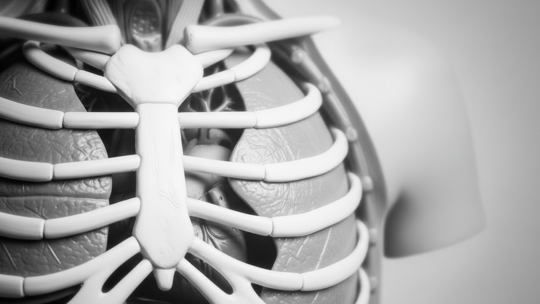
Heart
Model#: EZ28This 3D printed heart specimen preserves superficial cardiac anatomy and the bases of the great vessels. All four chambers (atria and ventricles) are preserved, with the pericardial reflections on the left atrium demarcating the position...
Right thoracic wall - axilla, and the root of the neck
Model#: EZ12This 3D printed specimen preserves a dissection of the right thoracic wall, axilla, and the root of the neck. The specimen is cut just parasagittally and the visceral contents of the chest have been removed....
Posterior Body Wall / Ventral Deep Dissection
Model#: EZ4This 3D printed specimen complements our dorsal dissection specimen (MP1400) by presenting a ventral deep dissection of axial anatomy from the head, neck, axillae, thorax, and abdomen to the proximal portion of the thighs. The...
Nervous System Dissection (posterior view)
Model#: EZ3This 3D printed specimen presents a unique view of axial anatomy, presenting a dorsal deep dissection of the head, neck, axillae, thorax, abdomen, and gluteal regions. The removal of the posterior portions of the cranium...
Posterior Abdominal wall
Model#: EZ2This large 3D-printed specimen displays the entire male posterior abdominal wall from the diaphragm to the pelvic brim, as well as pelvic anatomy and to the proximal thigh. This same individual specimen is also available...
Bowel - Portion of Jejenum
Model#: EZ32This 3D printed specimen presents a small loop of jejenum and mesentery. A window into the mesentery, fat and visceral peritoneum has been removed to illustrate the arterial arcades in the mesentery.
Bowel - Portion of Ileum
Model#: EZ31This 3D printed specimen demonstrates a small loop of ileum and mesentery. A window into the mesentery has been dissected (removing fat and visceral peritoneum) to show arterial arcades in the mesentery.
Bronchial Tree
Model#: EZ27This 3D printed specimen presents the conducting pathways of the respiratory system from the trachea, carina, and complete right and left bronchial trees to the level of the tertiary lobar bronchi. Each set of lobar...


Erler-Zimmer 3D Torso and Organ Models for Comprehensive Anatomical Study
Erler-Zimmer 3D torso and organ models are designed to support structured anatomical education by providing detailed representations of human body systems and internal organs. These models allow learners to explore anatomical relationships within the torso, offering clear insight into how major systems are organized and function together.
Developed for academic and clinical learning environments, these models support both foundational and advanced instruction by presenting complex anatomical structures in a clear, accessible format.
Designed for System-Level Visualization and Structural Clarity
Erler-Zimmer models emphasize accurate spatial representation and detailed structure, allowing users to study the positioning and interaction of organs within the human body. Their three-dimensional design supports a deeper understanding of anatomical organization across multiple systems.
Key Features of Erler-Zimmer 3D Torso and Organ Models
- Comprehensive representation of major organ systems
- Three-dimensional structure for spatial understanding
- Removable and sectional components for focused study
- Detailed visualization of internal anatomical relationships
- Realistic proportions and anatomical positioning
- Durable construction for repeated educational use
These features enable educators to demonstrate how different systems interact within the torso while helping learners identify and understand individual organs and structures.
Built for Anatomy Education and Clinical Training Environments
Erler-Zimmer 3D torso and organ models are used in settings where full-body system understanding and internal anatomy are studied. Their design supports both instructor-led demonstrations and independent learning.
Common Educational Applications
- University anatomy and medical programs
- Nursing and allied health education
- Clinical and healthcare training environments
- Biology and life science instruction
- Advanced anatomical study and demonstration
These models provide consistent support for teaching system-level anatomy and internal structure relationships within the human body.
Supporting Integrated Understanding of Human Body Systems
Erler-Zimmer 3D torso and organ models help learners understand how multiple organ systems function together within the body. By presenting anatomical structures in a cohesive and spatially accurate format, these models support deeper comprehension of physiological organization.
Whether used for demonstration, structured coursework, or advanced study, Erler-Zimmer 3D torso and organ models provide reliable tools for effective and accurate anatomical education.


