


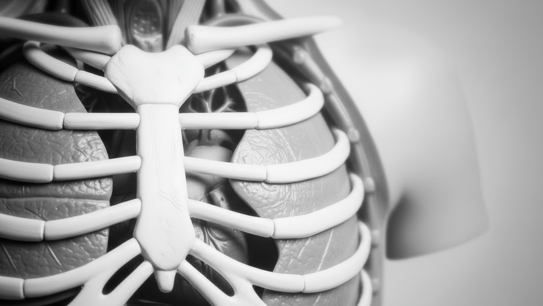
Erler-Zimmer 3D Extremity Models (23)
Erler-Zimmer 3D extremity models are designed to support structured instruction of upper and lower limb anatomy across classroom, collegiate, and clinical learning environments. This collection includes detailed three-dimensional teaching models that illustrate bones, muscles, ligaments, and functional movement of arms, hands, legs, and feet. These models support clear demonstration, hands-on learning, and system-level study for anatomy and healthcare education.
Foot - Deep plantar structures
Institutional List Price: $0.00Model#: EZ56This 3D printed specimen provides a view of deep plantar structures of a right foot. Medially, the cut edge of the great saphenous vein is visible within the superficial fascia, just anterior to the cut...
Foot - Superficial and deep structures of the distal leg and foot
Institutional List Price: $0.00Model#: EZ55This 3D printed specimen presents both superficial and deep structures of a right distal leg and foot. Proximally, the posterior compartment of the leg has been dissected to remove the triceps surae muscles and tendocalcaneous...
Foot - Superficial and deep dissection of distal leg and foot
Institutional List Price: $0.00Model#: EZ54This 3D printed specimens preserves a mixed superficial and deep dissection of a left distal leg and foot. Posteriorly, the compartment muscles and neurovascular structures have been removed to isolate the tendocalcaneous and expose the...
Foot - Plantar surface & superficial dissection on the dorsum
Institutional List Price: $0.00Model#: EZ53This 3D printed specimen is a left foot with superficial structures exposed on the dorsum, and the superficial layer of muscles and nerves on the plantar surface. The anterior portion of the plantar aponeurosis has...
Foot - Structures of the plantar surface
Institutional List Price: $0.00Model#: EZ52This 3D print records the anatomy of a right distal leg and the deep structures of the plantar surface of the foot. Proximally, the tibia, fibula, interosseous membrane, and leg muscles are discernable in cross-section....
Popliteal Fossa
Institutional List Price: $0.00Model#: EZ50This 3D printed specimen preserves the distal thigh and proximal leg, dissected posteriorly to demonstrate the contents of the popliteal fossa and surrounding region. The proximal cross-section demonstrates the anterior, posterior and medial compartment muscles,...
Popliteal Fossa distal thigh and proximal leg
Institutional List Price: $0.00Model#: EZ49This 3D printed specimen preserves the distal thigh and proximal leg, dissected posteriorly to demonstrate the contents of the popliteal fossa and surrounding region.
Lower limb G superficial dissection
Institutional List Price: $0.00Model#: EZ47This 3D printed specimen represents the remainder of the lower limb portions of our male abdominopelvic and proximal thigh specimen (MP1765), sectioned proximally near midthigh and continuous to the partially dissected foot. The transverse section...
Lower Limb superficial veins
Institutional List Price: $0.00Model#: EZ46This 3D printed specimen presents a superficial dissection of a left lower limb, from just proximal to the knee joint to a complete foot. The skin and superficial fascia have been removed to display the...
Lower Limb Musculature
Institutional List Price: $0.00Model#: EZ44This 3D printed specimen preserves a superficial dissection of the lower limb musculature from the mid-thigh to mid-leg, as well as nerves and vessels of the popliteal fossa. The insertions of the muscles of the...
Lower Limb - deep dissection
Institutional List Price: $0.00Model#: EZ43This 3D printed specimen consists of a right partial lower limb sectioned just proximal to the knee joint and complete through a partially dissected foot exposing the structures on the dorsum.
Cubital fossa - muscles, large nerves and the brachial artery
Institutional List Price: $0.00Model#: EZ34This 3D printed specimen presents a left distal arm and proximal forearm with all skin, subcutaneous fat and superficial cutaneous nerves and veins removed. The elbow region partially flexed to display the arrangement of muscles...
Cubital Fossa
Institutional List Price: $0.00Model#: EZ33This 3D printed cubital fossa displays a superficial dissection of the right distal arm and proximal forearm. The skin and superficial fascia has been removed anteriorly, medially and laterally to expose the superficial veins (basilic,...
Hand
Institutional List Price: $0.00Model#: EZ16This 3D printed specimen demonstrates a superficial dissection of a left hand and wrist. Anteriorly, the transverse carpal and palmar carpal ligaments have been removed to expose the tendons and nerves traversing the carpal tunnel...
Upper Limb - biceps, bones and ligaments
Institutional List Price: $0.00Model#: EZ10This 3D-printed specimen shows the origin and insertion of biceps (most other arm and shoulder muscle bellies have been removed). The long head of biceps arises from the supraglenoid tubercle (hidden from view) and travels...
Forearm and hand - deep dissection
Institutional List Price: $0.00Model#: EZ9This 3D printed specimen of a left upper limb preserves a deep dissection from the distal humerus to the palmar surface of hand.
Deep upper limb and hand
Institutional List Price: $0.00Model#: EZ8This 3D print of a superficially dissected right upper limb specimen displays a mixture of the vascular, nervous and muscular anatomy of the distal arm, forearm and hand.
Forearm and hand - superficial and deep dissection
Institutional List Price: $0.00Model#: EZ7This 3D printed specimen preserves a mixed superficial and deep dissection of the anterior aspect of a right distal arm, forearm and hand.
Upper Limb - elbow, forearm and hand
Institutional List Price: $0.00Model#: EZ6This 3D-printed specimen displays a great deal of upper limb anatomy. In the distal arm and elbow/ cubital fossa region it shows the arrangement of the biceps tendon, brachial artery and median nerve arranged from...
Upper Limb
Institutional List Price: $0.00Model#: EZ5This 3D-printed specimen demonstrates the superficial anatomy of a left upper limb from the blade of the scapula to the hand. The skin and superficial and deep fascia has been removed from most of the...
Foot - Parasagittal cross-section
Institutional List Price: $342.00Model#: EZ51This 3D printed specimen provides a parasagittal cross-section through the medial aspect of the right distal tibia and foot, displaying the skeletal structures of the medial longitudinal arch of the foot and surrounding soft-tissue structures.
Lower limb G superficial dissection with male left pelvis
Institutional List Price: $9,195.00Model#: EZ48This 3D printed specimen combines the Lower limb G superficial dissection (Ref.no. MP1816) with the male left pelvis (Ref.no. MP1765).
Upper Limb Ligaments
Institutional List Price: $1,494.00Model#: EZ11This 3D printed specimen presents the entire upper limb skeleton and ligaments from the pectoral girdle to the hand.


Erler-Zimmer 3D Extremity Models for Limb Anatomy and Movement Study
Erler-Zimmer 3D extremity models are designed to support structured anatomical education focused on the upper and lower limbs. These models provide detailed representations of bones, joints, muscles, and connective structures, allowing learners to study movement, alignment, and functional anatomy with clarity.
Developed for academic and clinical training environments, these models help users explore how limb structures interact to support mobility, stability, and coordination across different regions of the body.
Designed for Functional and Structural Understanding of Limbs
Erler-Zimmer extremity models emphasize accurate anatomical positioning and joint articulation, enabling clear visualization of how bones and soft tissues work together. Their three-dimensional design supports both static study and movement-based analysis.
Key Features of Erler-Zimmer 3D Extremity Models
- Detailed representation of upper and lower limb anatomy
- Three-dimensional structure for spatial and functional understanding
- Articulated joints to demonstrate movement and range of motion
- Visualization of bones, ligaments, and muscle structures
- Accurate anatomical proportions and alignment
- Durable construction for repeated educational use
These features allow educators to demonstrate how limb structures function together while helping learners understand movement mechanics and anatomical relationships.
Built for Anatomy, Rehabilitation, and Clinical Education
Erler-Zimmer 3D extremity models are used in learning environments where limb structure and function are central to instruction. Their design supports both guided teaching and independent anatomical exploration.
Common Educational Applications
- University anatomy and medical programs
- Physical therapy and rehabilitation education
- Sports science and kinesiology programs
- Orthopedic and clinical training environments
- Advanced biology and human movement studies
These models provide consistent support for teaching joint mechanics, structural alignment, and functional movement within the limbs.
Supporting Movement-Based and Functional Anatomy Learning
Erler-Zimmer 3D extremity models help learners understand how individual components of the limbs contribute to overall motion and stability. By presenting anatomy in a functional context, these models support deeper comprehension of coordination, load distribution, and biomechanical relationships.
Whether used for demonstration, structured coursework, or advanced study, Erler-Zimmer 3D extremity models provide reliable tools for teaching limb anatomy and movement in a clear and engaging way.


