


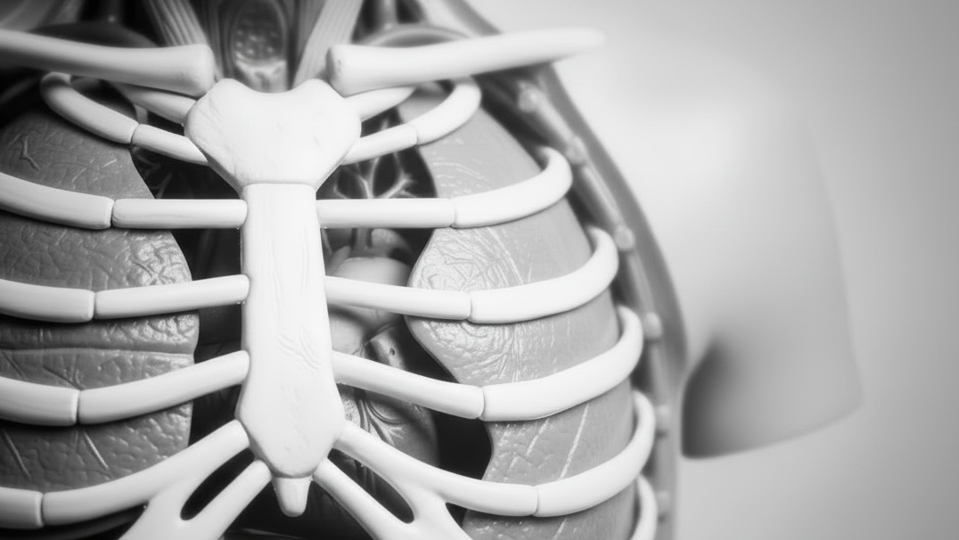
Erler-Zimmer 3D Joint Models (6)
Erler-Zimmer 3D joint models are designed to support structured instruction of joint anatomy and movement mechanics across classroom, collegiate, and clinical learning environments. This collection includes detailed three-dimensional teaching models that illustrate articulating bones, ligaments, cartilage, and functional joint relationships. These models support clear demonstration, hands-on learning, and system-level study for anatomy and healthcare education.
Knee Joint, extended
Institutional List Price: $0.00Model#: EZ41This 3D printed specimen demonstrates the ligaments of the knee joint with the leg in extension, it represents the same specimen as MP1800 knee joint printed in a flexed position. Both tibial and fibular collateral...
Shoulder - deep dissection of a right shoulder girdle
Institutional List Price: $0.00Model#: EZ15This 3D printed specimen preserves a deep dissection of a right shoulder girdle, consisting of a complete scapula, lateral clavicle, and proximal humerus. In the anterior view, the subscapularis muscle is present but sectioned to...
Flexed knee joint deep dissection
Institutional List Price: $0.00Model#: EZ42This 3D printed specimen displays a deep dissection of a left knee joint with the internal joint capsule structures relative to superficial tissues in a flexed position.
Knee Joint, flexed
Institutional List Price: $0.00Model#: EZ40This 3D printed specimen demonstrates the ligaments of the knee joint with the leg in flexion. In the anterior view, with the patella and part of the patellar ligament removed, the medial and lateral menisci...
Shoulder - deep dissection of the left shoulder joint, musculature, and associated nerves and vessels
Institutional List Price: $0.00Model#: EZ14This 3D printed specimen presents a deep dissection of the left shoulder joint, musculature, and associated nerves and vessels of the scapula and proximal humerus (to near midshaft). Anteriorly, the deltoid muscle has been detached...
Shoulder (left) - Superficial muscles and axillary/brachial artery
Institutional List Price: $0.00Model#: EZ13This printed 3D left shoulder specimen consists of the scapula, humerus (sectioned near midshaft) and clavicle (sectioned at midshaft) with the superficial muscles around the shoulder joint, the rotator cuff muscles and the axillary artery...


Erler-Zimmer 3D Joint Models for Articulation and Movement-Based Anatomy Education
Erler-Zimmer 3D joint models are designed to support structured anatomical education focused on joint structure, articulation, and movement. These models provide detailed representations of major joints in the human body, allowing learners to study how bones, ligaments, and surrounding structures interact to produce motion.
Developed for academic and clinical training environments, these models help users explore joint mechanics, alignment, and functional relationships with clarity and precision.
Designed for Clear Understanding of Joint Structure and Function
Erler-Zimmer joint models emphasize anatomical accuracy and functional movement, enabling learners to visualize how joints operate in real-world conditions. Their three-dimensional design supports both static study and dynamic demonstration.
Key Features of Erler-Zimmer 3D Joint Models
- Accurate representation of major human joints
- Articulated components to demonstrate natural movement
- Visualization of ligaments and structural support elements
- Three-dimensional design for spatial understanding
- Realistic anatomical positioning and alignment
- Durable construction for repeated instructional use
These features allow educators to demonstrate joint mechanics clearly while helping learners understand how structure supports movement and stability.
Built for Anatomy, Rehabilitation, and Clinical Training Environments
Erler-Zimmer 3D joint models are used in educational settings where joint function and movement are central to instruction. Their design supports both instructor-led demonstrations and hands-on exploration.
Common Educational Applications
- University anatomy and medical programs
- Physical therapy and rehabilitation education
- Sports science and kinesiology programs
- Orthopedic and clinical training environments
- Advanced biology and human movement studies
These models provide consistent support for teaching articulation, joint stability, and movement patterns within the human body.
Supporting Functional and Movement-Based Learning
Erler-Zimmer 3D joint models help learners understand how joints enable movement while maintaining structural support. By presenting anatomy in a functional context, these models support deeper comprehension of motion, flexibility, and mechanical interaction between structures.
Whether used for demonstration, structured coursework, or advanced study, Erler-Zimmer 3D joint models provide reliable tools for teaching joint anatomy and movement in a clear and engaging way.


