


Erler-Zimmer 3D Head and Neck Models (11)
Erler-Zimmer 3D head and neck models are designed to support structured instruction of regional anatomy across classroom, collegiate, and clinical learning environments. This collection includes detailed three-dimensional teaching models that illustrate cranial structures, facial anatomy, musculature, glands, and airway components. These models enable clear visualization of anatomical relationships and support demonstration, hands-on learning, and system-level study for anatomy and healthcare education.
Medial Orbit
Institutional List Price: $0.00Model#: EZ26This 3D print displays the orbital contents and its close relations as viewed from the medial perspective when the majority of the lateral wall of the nasal cavity and the intervening ethmoidal sinuses have been...
Superior Orbit
Institutional List Price: $0.00Model#: EZ24This 3D printed model captures a dissection in which the calvaria and cerebrum have been removed to expose the floors of the anterior and middle cranial fossae. The midbrain has been sectioned at the level...
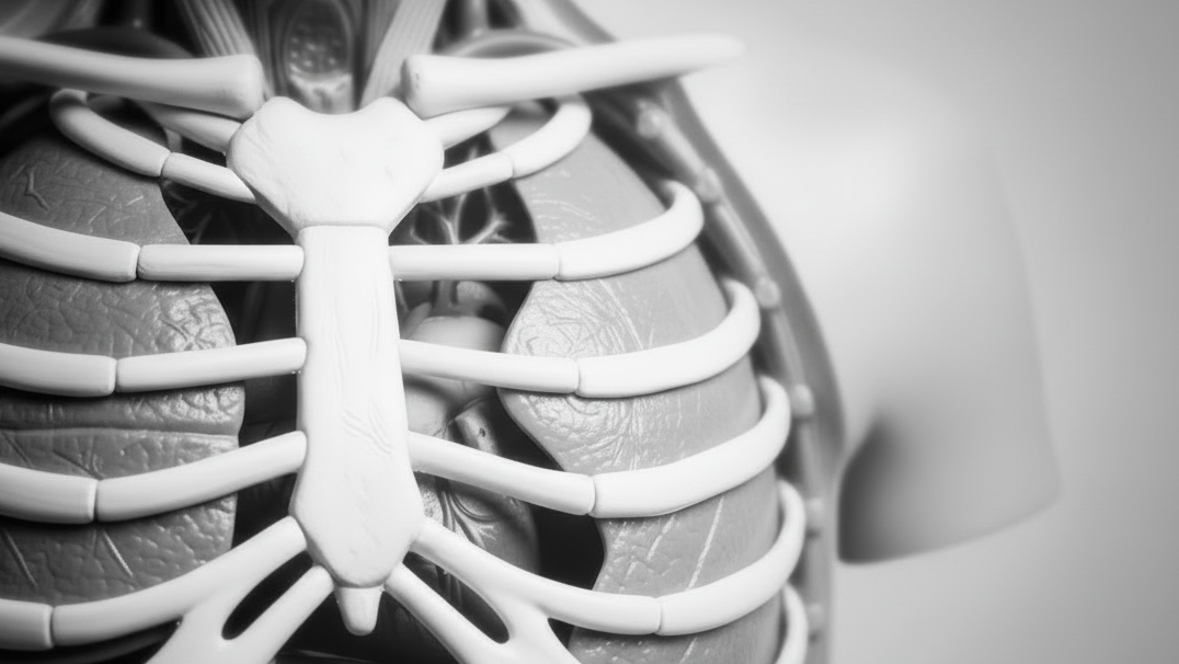
Head and visceral column of the neck
Institutional List Price: $0.00Model#: EZ23This 3D print specimen preserves a series of features of the head and visceral column of the neck: The face: On the right side of the head the parotid gland has been removed to reveal...
Deep face/Infratemporal fossa
Institutional List Price: $0.00Model#: EZ22In this 3D printed specimen of a midsagittally-sectioned right face and neck, the ramus, coronoid process and head of the mandible have been removed to expose the deep part of the infratemporal fossa. The pterygoid...
Head and Neck
Institutional List Price: $0.00Model#: EZ21This 3D printed specimen of a parasagittally sectioned head and neck demonstrates a range of anatomical features: Lateral aspect of the face: A window has been created to expose the parotid region. The pinna of...
Temporal Bone Model, Set of 3
Institutional List Price: $0.00Model#: EZ19This 3 part 3D printed model derived from CT data highlights the complex anatomy of the temporal bone including bone ossicles, canals, chambers, foramina and air spaces. In addition, the spatial relations between temporal bone...
Dural Skull
Institutional List Price: $0.00Model#: EZ18This 3D print of a dissected and opened cranial cavity displays the dural folds and dural venous sinuses, including the falx cerebri (preserved by a retained midsagittal portion of the calvaria. The intact tentorium cerebelli...
Circle of Willis
Institutional List Price: $0.00Model#: EZ17This 3D printed specimen demonstrates the intracranial arteries that supply the brain relative to the inferior portions of the viscero- and neurocranium. This print was created by careful segmentation of angiographic data. The model shows...
Head, Neck and Shoulder with angiosomes
Institutional List Price: $0.00Model#: EZ1This large 3D printed specimen displays a great deal of anatomy spanning the head, neck, thorax, axillae and upper limbs.
Lateral Orbit
Institutional List Price: $348.00Model#: EZ25This 3D printed specimen shows the orbit from the lateral perspective when the bony lateral wall and part of the calvaria of the skull have been removed. The frontal and temporal lobes of the brain...
Paranasal Sinus model
Institutional List Price: $551.00Model#: EZ20This unique model has been created from CT imaging and segmentation of the internal spaces of the viscerocranium. Parts of the skull have been retained but sections or windows have been removed to expose the...


Erler-Zimmer 3D Head and Neck Models for Advanced Anatomical Visualization
Erler-Zimmer 3D head and neck models are designed to support structured anatomical education, providing detailed representations of cranial, facial, and cervical structures. These models enable clear visualization of complex anatomical relationships within the head and neck region, supporting both foundational and advanced study.
Developed for academic and clinical learning environments, these models help learners explore anatomical organization, structural positioning, and system interactions with clarity and precision.
Designed for Detailed Study of Head and Neck Anatomy
Erler-Zimmer models emphasize spatial accuracy and structural clarity, allowing users to examine both external features and internal anatomical components. Their three-dimensional design supports deeper understanding of anatomical layering and regional relationships.
Key Features of Erler-Zimmer 3D Head and Neck Models
- Accurate representation of cranial, facial, and cervical anatomy
- Three-dimensional structure for spatial understanding
- Detailed visualization of muscles, nerves, and vascular systems
- Sectional and layered components for focused study
- Realistic anatomical proportions and positioning
- Durable construction for repeated educational use
These features allow educators to present complex anatomical structures in a clear and organized format while helping learners build confidence in identifying key components of the head and neck.
Built for Anatomy Education and Clinical Training Environments
Erler-Zimmer 3D head and neck models are used in educational settings where detailed anatomical study and regional understanding are required. Their design supports both demonstration-based teaching and independent exploration.
Common Educational Applications
- University anatomy and medical programs
- Dental and craniofacial studies
- Nursing and allied health education
- Clinical and surgical training environments
- Advanced biology and anatomical instruction
These models provide consistent support for teaching anatomical relationships within the head and neck, helping learners connect structure with function.
Supporting Spatial and System-Level Understanding
Erler-Zimmer 3D head and neck models help learners understand how different anatomical systems interact within a confined and complex region of the body. By presenting structures in three dimensions, these models enhance comprehension of positioning, depth, and connectivity.
Whether used for demonstration, structured coursework, or advanced study, Erler-Zimmer 3D head and neck models provide reliable tools for accurate and effective anatomical education.


