


Technology has transformed nearly every aspect of education, and medical training is no exception. Today's students have access to advanced learning tools such as virtual reality (VR), augmented reality (AR), 3D anatomy software, digital simulations, and online learning platforms. These innovations have expanded educational opportunities and improved access to anatomical knowledge.
However, despite the rapid growth of digital learning technologies, hands-on anatomy learning remains an essential component of effective medical education. Physical anatomical models continue to play a critical role in helping students understand complex anatomical structures, develop clinical skills, and retain knowledge more effectively.
Anatomy education has evolved significantly over the centuries.
Traditional teaching methods relied heavily on textbooks, lectures, cadaver dissection, and physical anatomical models. Today, educators have access to sophisticated digital tools that allow students to explore the human body in remarkable detail.
Some of the most common digital anatomy resources include:
While these technologies provide valuable learning opportunities, they do not fully replace the educational benefits of physical interaction with anatomical structures.
The most effective anatomy programs often combine digital tools with hands-on learning experiences.
Research in educational psychology consistently shows that active learning produces better outcomes than passive learning.
When students physically interact with anatomical models, they engage multiple senses simultaneously, including:
This multisensory approach helps strengthen neural connections and improves understanding of anatomical relationships.
For example, rotating a skeletal model or removing organs from a torso model allows students to actively explore anatomy rather than simply viewing it on a screen.
This level of engagement often leads to deeper comprehension and stronger long-term retention.
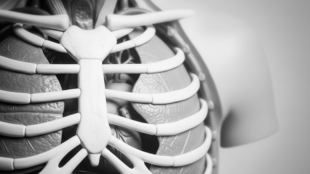
Human anatomy is inherently three-dimensional.
Students must understand how structures relate to one another in space, including:
While digital platforms can simulate three-dimensional anatomy, physical models provide a tangible experience that many students find easier to understand.
Being able to hold, examine, and manipulate anatomical structures from different angles helps learners build accurate mental representations of the human body.
Digital learning tools offer tremendous advantages, but they also have limitations.
Viewing anatomy on a screen does not provide the same sensory experience as handling a physical model.
Students may struggle to fully appreciate:
Without tactile interaction, some learners may find it more difficult to develop a complete understanding of anatomical structures.
Modern students spend significant amounts of time looking at screens.
Between lectures, assignments, online resources, and digital anatomy platforms, prolonged screen exposure can contribute to:
Hands-on learning activities provide a welcome alternative that can improve focus and reinforce key concepts through active participation.
Digital tools depend on hardware, software, and internet connectivity.
Educational institutions may face challenges such as:
Physical anatomical models provide a reliable teaching solution that requires no software updates or technical infrastructure.
Healthcare students must eventually apply their anatomical knowledge in clinical settings.
Physical anatomical models help bridge the gap between classroom learning and real-world practice by allowing students to:
This hands-on experience builds confidence and prepares students for future patient care responsibilities.
Successful healthcare professionals must do more than memorize anatomy.
They must understand how anatomical structures function together and how diseases, injuries, and treatments affect the body.
Physical models encourage students to ask questions, explore relationships, and develop critical thinking skills that support clinical decision-making.
This deeper level of understanding often translates into improved performance during clinical training.
Even in highly digital learning environments, anatomical models remain a cornerstone of anatomy instruction.
Educators value physical models because they:
Anatomical models also provide consistency across educational settings, from high school STEM programs to medical schools and healthcare training institutions.
The question is no longer whether educators should choose physical models or digital tools.
Instead, the most effective approach is integrating both.
A blended learning strategy allows students to benefit from:
Together, these tools create a comprehensive educational experience that supports both foundational knowledge and clinical skill development.
To maximize learning outcomes, educators should invest in high-quality anatomical models that align with their curriculum objectives.
Popular options include:
Well-designed models provide accurate anatomical detail and help students connect theoretical concepts with real-world healthcare applications.
While digital technologies have revolutionized anatomy education, hands-on learning remains as important as ever. Physical anatomical models offer unique educational benefits that digital tools alone cannot fully replicate.
By promoting active participation, enhancing spatial understanding, improving knowledge retention, and supporting clinical readiness, hands-on anatomy learning continues to play a vital role in healthcare education.
The future of anatomy education is not about replacing traditional learning methods it is about combining the strengths of both physical and digital tools to create richer, more effective learning experiences for the next generation of healthcare professionals.
For educators seeking to improve student engagement and learning outcomes, anatomical models remain one of the most valuable teaching resources available today.
Serving Institutions Nationwide: Holt Anatomical supports nursing, anatomy, and biology programs across the United States. Our team works with faculty, lab managers, and purchasing departments to provide accurate models, reliable fulfillment, and long-term support.


