


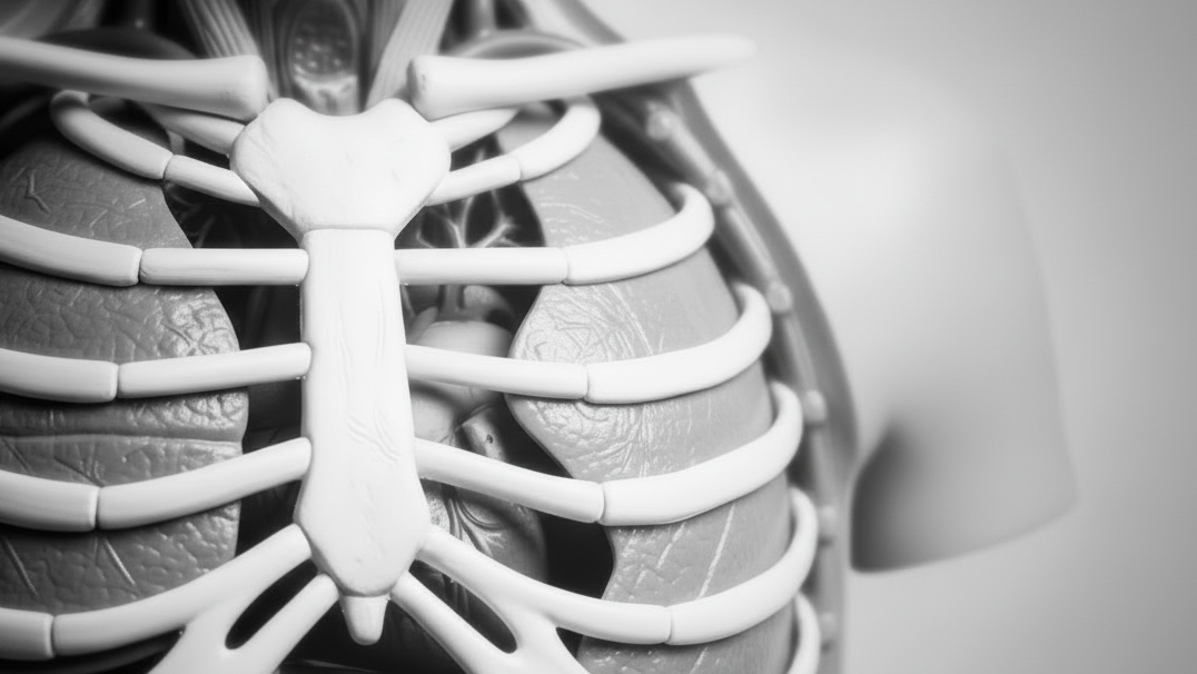
Erler-Zimmer Models (56)
Erler-Zimmer models are designed to support structured anatomical and healthcare education across classroom, collegiate, and clinical learning environments. This collection includes detailed teaching models representing human anatomy, organs, and physiological systems. Built for clear demonstration and hands-on study, these models support anatomy instruction, medical training, and healthcare education programs.
Knee Joint, extended
Institutional List Price: $0.00Model#: EZ41This 3D printed specimen demonstrates the ligaments of the knee joint with the leg in extension, it represents the same specimen as MP1800 knee joint printed in a flexed position. Both tibial and fibular collateral...
Foot - Deep plantar structures
Institutional List Price: $0.00Model#: EZ56This 3D printed specimen provides a view of deep plantar structures of a right foot. Medially, the cut edge of the great saphenous vein is visible within the superficial fascia, just anterior to the cut...
Shoulder - deep dissection of a right shoulder girdle
Institutional List Price: $0.00Model#: EZ15This 3D printed specimen preserves a deep dissection of a right shoulder girdle, consisting of a complete scapula, lateral clavicle, and proximal humerus. In the anterior view, the subscapularis muscle is present but sectioned to...
Foot - Superficial and deep structures of the distal leg and foot
Institutional List Price: $0.00Model#: EZ55This 3D printed specimen presents both superficial and deep structures of a right distal leg and foot. Proximally, the posterior compartment of the leg has been dissected to remove the triceps surae muscles and tendocalcaneous...
Foot - Superficial and deep dissection of distal leg and foot
Institutional List Price: $0.00Model#: EZ54This 3D printed specimens preserves a mixed superficial and deep dissection of a left distal leg and foot. Posteriorly, the compartment muscles and neurovascular structures have been removed to isolate the tendocalcaneous and expose the...
Foot - Plantar surface & superficial dissection on the dorsum
Institutional List Price: $0.00Model#: EZ53This 3D printed specimen is a left foot with superficial structures exposed on the dorsum, and the superficial layer of muscles and nerves on the plantar surface. The anterior portion of the plantar aponeurosis has...
Foot - Structures of the plantar surface
Institutional List Price: $0.00Model#: EZ52This 3D print records the anatomy of a right distal leg and the deep structures of the plantar surface of the foot. Proximally, the tibia, fibula, interosseous membrane, and leg muscles are discernable in cross-section....
Popliteal Fossa
Institutional List Price: $0.00Model#: EZ50This 3D printed specimen preserves the distal thigh and proximal leg, dissected posteriorly to demonstrate the contents of the popliteal fossa and surrounding region. The proximal cross-section demonstrates the anterior, posterior and medial compartment muscles,...
Popliteal Fossa distal thigh and proximal leg
Institutional List Price: $0.00Model#: EZ49This 3D printed specimen preserves the distal thigh and proximal leg, dissected posteriorly to demonstrate the contents of the popliteal fossa and surrounding region.
Lower limb G superficial dissection
Institutional List Price: $0.00Model#: EZ47This 3D printed specimen represents the remainder of the lower limb portions of our male abdominopelvic and proximal thigh specimen (MP1765), sectioned proximally near midthigh and continuous to the partially dissected foot. The transverse section...
Lower Limb superficial veins
Institutional List Price: $0.00Model#: EZ46This 3D printed specimen presents a superficial dissection of a left lower limb, from just proximal to the knee joint to a complete foot. The skin and superficial fascia have been removed to display the...
Lower Limb - deep dissection of a left pelvis and thigh
Institutional List Price: $0.00Model#: EZ45This 3D printed specimen presents a deep dissection of a left pelvis and thigh to show the course of the femoral artery and sciatic nerve from their proximal origins to the midshaft of the femur....
Lower Limb Musculature
Institutional List Price: $0.00Model#: EZ44This 3D printed specimen preserves a superficial dissection of the lower limb musculature from the mid-thigh to mid-leg, as well as nerves and vessels of the popliteal fossa. The insertions of the muscles of the...
Lower Limb - deep dissection
Institutional List Price: $0.00Model#: EZ43This 3D printed specimen consists of a right partial lower limb sectioned just proximal to the knee joint and complete through a partially dissected foot exposing the structures on the dorsum.
Flexed knee joint deep dissection
Institutional List Price: $0.00Model#: EZ42This 3D printed specimen displays a deep dissection of a left knee joint with the internal joint capsule structures relative to superficial tissues in a flexed position.
Knee Joint, flexed
Institutional List Price: $0.00Model#: EZ40This 3D printed specimen demonstrates the ligaments of the knee joint with the leg in flexion. In the anterior view, with the patella and part of the patellar ligament removed, the medial and lateral menisci...
Female right pelvis
Institutional List Price: $0.00Model#: EZ39This 3D printed specimen represents a female right pelvis, sectioned along the midsagittal plane and transversely across the level of the L4 vertebrae and the proximal thigh. The specimen has been dissected to demonstrate the...
Female left pelvis and proximal thigh
Institutional List Price: $0.00Model#: EZ37This 3D printed female left pelvis and proximal thigh preserves both superficial and deep structures of the true and false pelves, inguinal region, femoral triangle, and gluteal region. The specimen has been sectioned transversely through...
Male Pelvis
Institutional List Price: $0.00Model#: EZ36This multipart 3D printed specimen represents the inferior portions of our larger posterior abdominal wall print (MP1300) that displays the inferior posterior abdominal wall, the pelvic cavity and the proximal thigh (including the gluteal regions...
Male left pelvis and proximal thigh
Institutional List Price: $0.00Model#: EZ35This 3D printed male left pelvis and proximal thigh (sectioned through the midsagittal plane in the midline and transversely through the L3/4 intervertebral disc) shows superficial and deep structures of the true and false pelves,...
Cubital fossa - muscles, large nerves and the brachial artery
Institutional List Price: $0.00Model#: EZ34This 3D printed specimen presents a left distal arm and proximal forearm with all skin, subcutaneous fat and superficial cutaneous nerves and veins removed. The elbow region partially flexed to display the arrangement of muscles...
Cubital Fossa
Institutional List Price: $0.00Model#: EZ33This 3D printed cubital fossa displays a superficial dissection of the right distal arm and proximal forearm. The skin and superficial fascia has been removed anteriorly, medially and laterally to expose the superficial veins (basilic,...
Heart internal structures
Institutional List Price: $0.00Model#: EZ30This 3D printed heart has been dissected to display the internal structures of the chambers. At the base of the heart the termination of the superior vena cava is preserved entering the right atrium. Part...
Heart and the distal trachea, carina and primary bronchi
Institutional List Price: $0.00Model#: EZ29This 3D printed specimen preserves the external anatomy of the heart and the distal trachea, carina, and primary bronchi in the posterior mediastinum relative to the great vessels and left atrium (which demonstrates the pericardial...


