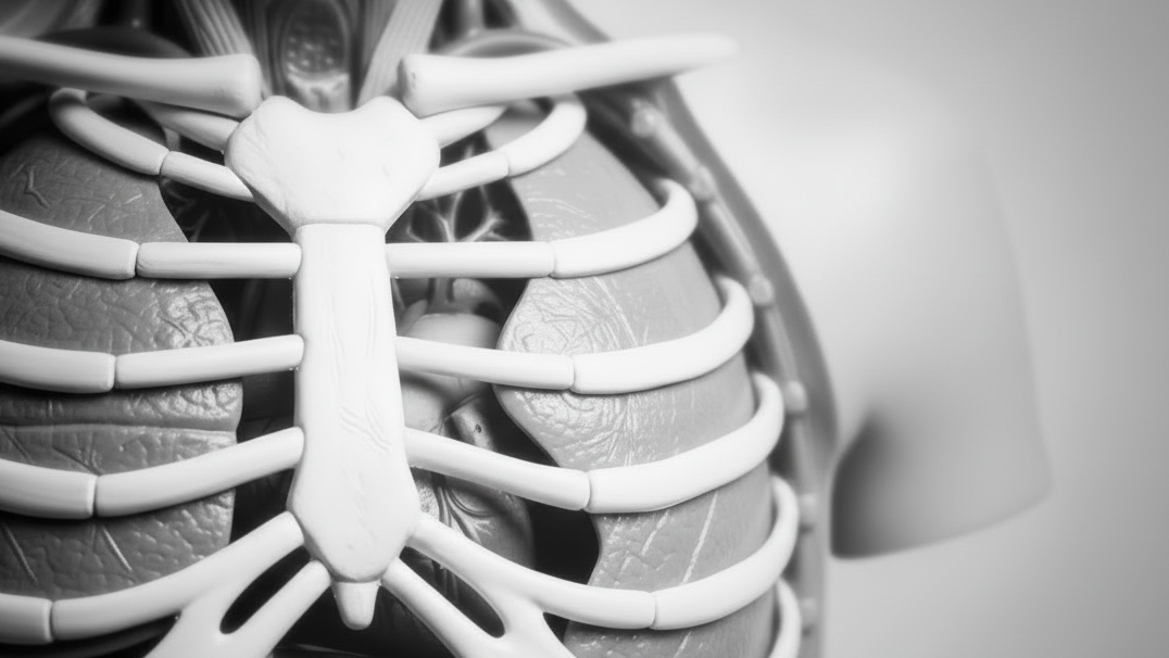
Human Torso Models for Anatomy and Medical Education
Human torso models provide a detailed, three-dimensional view of the major organs, muscles, blood vessels, nerves, and body systems located within the chest, abdomen, pelvis, head, and neck. Holt Anatomical offers educational torso models designed for anatomy classrooms, medical schools, nursing programs, biology courses, allied health education, laboratories, and clinical demonstrations.
This collection includes life-size human torso models, miniature desktop torsos, male and female torsos, unisex and dual-sex models, muscular torsos, open-back models, sectioned torsos, and advanced multi-part replicas with removable organs. Available products offer different levels of anatomical detail, scale, dissection, labeling, and removable components to support introductory through advanced instruction.
Study the Internal Organs of the Human Body
A human torso model helps learners understand the position, size, shape, and spatial relationships of the body’s major internal organs. Unlike a flat illustration, a three-dimensional model can be viewed from multiple angles and may allow organs to be removed for closer examination.
Depending on the model selected, students may be able to study the lungs, heart, liver, stomach, pancreas, spleen, kidneys, intestines, urinary bladder, reproductive organs, diaphragm, major blood vessels, brain, eye, and portions of the spinal column.
Removing and replacing organs can help students develop a clearer understanding of how the respiratory, cardiovascular, digestive, urinary, reproductive, nervous, and musculoskeletal systems are organized within the body.
Dissectible Torso Models with Removable Organs
Dissectible torso models contain removable organs and anatomical components that allow students to explore structures layer by layer. Learners can remove superficial organs, examine deeper structures, and reassemble the model in the correct anatomical arrangement.
Depending on the product, removable components may include the lungs, heart, liver, stomach, intestines, kidneys, bladder, brain, eye, reproductive organs, chest wall, abdominal wall, or sections of the back and spine.
The number of parts varies considerably among torso models. Basic educational torsos may contain fewer than ten removable components, while advanced models may separate into twenty, thirty, or more parts for detailed study.
Life-Size Human Torso Models
Life-size human torso models represent the upper body at or near natural scale. Their size allows instructors and students to examine organs and anatomical relationships in proportions similar to those of the human body.
These models are especially useful for instructor-led demonstrations, anatomy laboratories, medical and nursing programs, clinical education, and courses requiring detailed identification of internal structures.
Life-size torsos may include the head, neck, thorax, abdomen, and pelvis. Some models also include a removable muscular arm, open back, exposed spinal structures, or sex-specific reproductive anatomy.
Miniature and Desktop Torso Models
Miniature torso models provide a compact alternative to full-size anatomical torsos. They are useful for desktop study, small-group instruction, portable demonstrations, introductory biology courses, and classrooms with limited storage or display space.
Although reduced-size models may contain fewer removable parts or less surface detail than advanced life-size torsos, they can effectively demonstrate the general placement of the heart, lungs, liver, stomach, intestines, kidneys, and other major organs.
Review the dimensions and included components on each product page to determine whether a miniature model provides the level of detail required for your educational application.
Male and Female Human Torso Models
Male and female torso models allow students to study sex-specific reproductive and pelvic anatomy in the context of the complete body. A male torso may include structures such as the testes, penis, prostate, seminal vesicles, and associated urinary anatomy.
A female torso may include the uterus, ovaries, uterine tubes, vagina, mammary structures, and surrounding pelvic organs. The exact structures and number of removable components vary by product.
Sex-specific torso models are useful in anatomy, medicine, nursing, reproductive health, obstetrics, gynecology, urology, and patient education.
Unisex and Dual-Sex Torso Models
Unisex torso models focus primarily on structures common to the human body and may omit detailed external reproductive anatomy. They are practical for general anatomy, biology, nursing, and healthcare instruction where the primary focus is on the major internal organs and body systems.
Dual-sex torso models may include interchangeable male and female reproductive components. This allows one model to demonstrate both male and female pelvic anatomy while reducing the need to purchase two complete torsos.
Depending on the product, dual-sex inserts may be removable, interchangeable, sectioned, or shown in relationship to the bladder, rectum, blood vessels, and surrounding pelvic structures.
Open-Back Torso Models
Open-back torso models expose structures that are not visible from the front of a standard torso. Depending on the model, the open back may reveal the vertebral column, spinal cord, spinal nerve roots, ribs, kidneys, muscles, and posterior portions of internal organs.
These models help learners understand how the organs relate to the spine and posterior body wall. They may also provide a clearer view of the vertebral canal, intervertebral discs, spinal nerves, and deep muscular anatomy.
Open-back torsos are valuable for medical, nursing, physical therapy, chiropractic, radiology, and advanced anatomy education.
Open-Neck Torso Models
Open-neck torso models may expose the larynx, pharynx, trachea, esophagus, thyroid gland, cervical blood vessels, nerves, muscles, and upper portions of the respiratory and digestive systems.
These structures help students follow the airway and digestive tract from the head and neck into the thoracic cavity. Open-neck anatomy can also provide useful context for studying swallowing, speech, respiration, vascular pathways, and cervical organ relationships.
Muscular Torso Models
Muscular torso models combine internal organ anatomy with detailed representations of superficial and deep muscles. Depending on the product, muscles may be painted, fixed in place, or removable to expose underlying organs, blood vessels, nerves, bones, and deeper muscle layers.
These models may show the muscles of the chest, abdomen, back, shoulder, neck, and upper extremity. Some advanced torsos include a removable muscular arm for a more complete study of upper-body musculoskeletal anatomy.
Muscular torsos are useful for anatomy, medicine, physical therapy, occupational therapy, athletic training, kinesiology, massage therapy, and sports science education.
Torso Models with Removable Muscles
Models with removable muscles allow learners to study anatomy in layers. A superficial muscle may be removed to reveal deeper muscles, bones, nerves, blood vessels, or internal organs.
This approach supports dissection-style learning while providing a durable alternative to preserved specimens. Students can repeatedly remove and replace structures as they practice identification and develop an understanding of anatomical relationships.
The number and location of removable muscles vary by model. Review individual product specifications to determine which muscle groups and underlying structures are represented.
Torso Models with Head and Brain Anatomy
Some torso models include the head and portions of the brain, skull, eye, and cranial anatomy. The brain may separate into multiple pieces or be removable from the cranial cavity for closer examination.
Models with a head provide broader anatomical context by connecting the brain, airway, digestive tract, nerves, and blood vessels with the structures of the chest and abdomen.
Depending on the model, the head may also show facial muscles, the oral cavity, nasal cavity, eye, cranial nerves, or a sectioned view of internal structures.
Heart and Circulatory Anatomy in Torso Models
Most detailed torso models include a heart positioned between the lungs within the thoracic cavity. Depending on the product, the heart may be removable or divide into sections to reveal chambers, valves, and internal structures.
Torso models may also show the aorta, venae cavae, pulmonary vessels, carotid arteries, iliac vessels, and other major components of the circulatory system. These features help students understand how the heart and major blood vessels relate to surrounding organs.
Respiratory Anatomy in Human Torso Models
The respiratory structures represented on a torso model may include the nasal cavity, pharynx, larynx, trachea, bronchi, lungs, and diaphragm. Removable lungs allow learners to examine the heart, mediastinum, and deeper thoracic anatomy.
Some models divide the lungs into sections or show the bronchial tree, pulmonary vessels, and differences between the right and left lungs. These features support anatomy, respiratory therapy, nursing, medical, and biology education.
Digestive System Anatomy in Torso Models
Human torso models may show the digestive tract from the oral cavity and esophagus through the stomach, small intestine, large intestine, rectum, and anus. Accessory digestive organs may include the liver, gallbladder, pancreas, and salivary glands.
Removable digestive organs help students understand how structures are arranged within the abdominal cavity. Models may also show the duodenum, appendix, mesentery, peritoneum, blood vessels, or portions of the posterior abdominal wall.
Urinary System Anatomy in Torso Models
Urinary structures represented on torso models may include the kidneys, ureters, urinary bladder, and urethra. Some models show a kidney in section to reveal the cortex, medulla, renal pelvis, and internal collecting structures.
The urinary organs are often shown in relationship to surrounding digestive, reproductive, vascular, and skeletal structures. This helps learners understand the position of the kidneys behind the abdominal organs and the pathway of urine toward the bladder.
Reproductive Anatomy in Human Torso Models
Male, female, and dual-sex torso models may include reproductive structures within the pelvis. These models allow students to examine reproductive anatomy in relation to the urinary bladder, rectum, blood vessels, pelvic bones, and abdominal organs.
Depending on the model, reproductive components may be removable or sectioned to reveal internal anatomy. Interchangeable inserts may allow the same torso to demonstrate both male and female structures.
Sectional and Disc Torso Models
Sectional torso models divide the body into horizontal, sagittal, coronal, or other anatomical planes. These models help learners connect gross anatomy with cross-sectional views used in computed tomography, magnetic resonance imaging, and other medical imaging methods.
A disc torso may separate into a series of transverse slices that show organs, muscles, bones, blood vessels, and body cavities at different levels. Students can examine each section individually or assemble the slices to reconstruct the torso.
Sectional torsos are especially useful for radiology, medical imaging, surgery, anatomy, and advanced healthcare education.
Torso Models for Medical and Nursing Education
Medical and nursing programs use human torso models to teach organ identification, body systems, physical assessment, disease processes, surgical relationships, and clinical terminology. The ability to remove organs can support active learning and practical examination preparation.
Torso models provide a consistent reference for lectures, anatomy laboratories, simulation exercises, patient assessment instruction, and clinical demonstrations. Advanced models may support detailed study of nerves, blood vessels, muscles, reproductive anatomy, and sectional relationships.
Torso Models for Biology and Anatomy Classrooms
Biology and anatomy courses use torso models to introduce the organization of the human body and demonstrate how major organ systems interact. Students can follow the respiratory, cardiovascular, digestive, urinary, reproductive, and nervous systems within one integrated model.
Models with removable organs encourage hands-on participation and can be used for identification exercises, group activities, laboratory review, and practical examinations.
Torso Models for Allied Health Programs
Physical therapy, occupational therapy, respiratory therapy, radiologic technology, emergency medical, physician assistant, dental, and other allied health programs can use torso models to reinforce regional and system-based anatomy.
The most appropriate model depends on the program. Respiratory therapy courses may prioritize the airway and lungs, while radiology programs may benefit from sectional torsos. Therapy programs may prefer models that combine muscular, skeletal, nervous, and internal organ anatomy.
Human Torso Models for Patient Education
Healthcare professionals may use torso models to explain organ locations, medical conditions, diagnostic findings, surgical procedures, and treatment plans. Removable organs allow clinicians to show the position of a structure and its relationship to surrounding anatomy.
Torso models may support discussions involving heart or lung conditions, digestive disorders, kidney disease, reproductive health, surgery, cancer treatment, and other healthcare topics. Anatomical models are educational tools and are not intended to diagnose a medical condition.
How to Choose a Human Torso Model
The best torso model depends on the student level, structures being taught, available classroom space, and desired level of anatomical detail. A miniature torso may be appropriate for introductory courses or desktop study, while a life-size multi-part torso may be preferable for medical, nursing, or advanced anatomy programs.
Consider whether you need a male, female, unisex, or dual-sex model. You should also determine whether the torso should include a head, removable brain, open back, open neck, removable muscles, sectional anatomy, interchangeable reproductive organs, or a muscular arm.
The number of parts is another important consideration. Models with fewer components are generally easier to assemble and manage, while torsos with twenty or more parts provide greater detail for advanced study.
Review each Holt Anatomical product page for information about scale, model dimensions, sex, removable organs, open-back features, muscle anatomy, number of parts, mounting, and included educational materials.
Durable Torso Models for Repeated Instruction
Educational torso models provide a durable and consistent alternative to preserved anatomical specimens. They are designed for repeated handling during lectures, laboratory exercises, student review, practical examinations, clinical demonstrations, and patient consultations.
Browse Holt Anatomical’s collection of human torso models to compare life-size and miniature options, male and female anatomy, unisex and dual-sex models, removable organs, muscular structures, open backs, sectional anatomy, manufacturers, and availability. Choose the torso model that best supports your anatomy curriculum, medical program, nursing course, biology laboratory, allied health training, or patient education needs.